UW Research: Lily's Fund Fellows

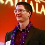
Antoine Madar, 2015 Fellow
Department of Neuroscience
We’re excited to have Antoine come on board as our third Lily’s Fund Fellow. Antoine is working in the neuroscience lab of Matt Jones, PhD and is interested in information processing in neural networks that are involved in memory and epilepsy. During his two-year fellowship, he will look for a correlation between the development of seizures and changes related to “pattern separation,” which is the essential neural process that allows us to recall distinctive memories of similar events. Antoine’s work aims to understand how abnormal activity in the hippocampus can lead both to seizures and memory impairments, in the hope of finding new treatments for both. Watch for updates.
During his two years as a Lily’s Fund research fellow, Antoine Madar has worked to better understand how a particular region of the brain affects both memory and epileptic seizures.
Our ability to remember events in our daily lives relies heavily on the hippocampus, a region of the brain that is also the starting point of many seizures. Indeed, the same characteristics that let the hippocampus recall memories make it vulnerable to seizures. To avoid this, a part of the hippocampus called the dentate gyrus filters excessive incoming signals. The breakdown of this gateway is a general feature of temporal lobe epilepsy.
“We know the dentate gyrus supports our ability to not confuse memories of similar events, but it is still unclear how it is able to do this. The first part of my project focused on understanding how the filtering properties of the dentate gyrus relate to its ability to avoid confusion of memories. I tested the hypothesis that the filtering role of the dentate gyrus is to transform incoming neural signals that are similar to each other into output signals that are less similar to each other, a process called pattern separation,” explains Antoine.
With his advisor Matt Jones and another previous student Laura Ewell, Antoine designed a novel experiment to measure pattern separation in brain slices from mice. They demonstrated for the first time that the dentate gyrus does, in fact, separate incoming neural signals at the level of single cells. This work is now complete and ready for publication. This refined understanding may help scientists design targeted treatments to restore normal filtering and pattern separation in the brain.
Antoine and his colleagues have started experiments to determine the effect of epilepsy on pattern separation and people’s ability to distinguish present experiences from similar ones in the past.
“I hypothesize that the development of epilepsy leads to impairments of pattern separation and thus to an increase in memory confusion. We have developed new behavioral tests to measure the ability of mice to distinguish between similar memories, and we are starting to use the test on mice at different stages of development of temporal lobe epilepsy,” says Antoine.
He hopes to establish the relationship between the advancement of epilepsy and impairments in pattern separation and memory. It is possible that pattern separation-related changes occur much earlier than more overt symptoms like seizures, and thus they could be used in the future as a tool for early diagnosis of seizure risk.
See Antoine’s full report with illustrations (PDF)
Acknowledgements: This is a collaborative project and Antoine wants to thank Jesse Pfammatter, Eli Wallace, Swetha Ravi, Rama Maganti and his advisor, Matt Jones.

Brandon Wright, 2013 Fellow
Brandon is a native of Louisiana, a UW grad and a PhD candidate working in physiology in the neuroscience lab of Dr. Meyer Jackson. In July of 2013, he became the second Lily’s Fund Fellow dedicating himself to cracking the code on epilepsy. He also has a personal stake in this game as his brother lives with epilepsy.
Seizures result from uncontrolled firing of neurons. Currently, electrodes and MRIs give us an idea where these electrical events take place but not an exact location. Brandon is using a technique called “voltage-imaging” to show where electrical pulses occur and where they will spread through the brain. Identifying the circuits or connections where runaway electrical activity occurs in the brain is the first step in finding the right treatment to stop the spread of seizures.
Brandon Wright, a PhD candidate working in the Neuroscience Department is working to better understand how electrical events happen in the brain. Brandon is working with slices of brain tissue, subjecting each to low-frequency electrical stimulation. The goal is to record the responses under normal conditions, then compare these results to slices that have been induced to a hyperactive state, or those where anti-seizure drugs have been introduced. He looks at how and where the signal spreads from the site of origin. This kind of basic research helps us gain a better understanding of how seizures take hold in the brain. Brandon’s fellowship continues through mid-2014. A call has gone out for the 2014-2015 Lily’s Fund fellow.
Our second Lily’s Fund fellow, Brandon Wright, has been studying the circuitry within the hippocampal dentate gyrus, a structure toward the back of the brain that becomes intensely active during a common form of epilepsy called temporal lobe epilepsy. The goal of Brandon’s work is to show that these natural brain circuits are involved in processes that contribute to learning and memory, as well as epilepsy.
Neural circuitry is plastic, meaning the connections can be modified by experience (such as learning) or by pathological events (such as seizures).
The connections between nerve cells — the synapses — can be strengthened using intense electrical signals, a process called long-term potentiation. It is thought that long-term potentiation is a required step in the formation of memories. No long-term potentiation, no learning new things.
Brandon is inducing long-term potentiation in the circuitry of the dentate gyrus by applying a series of high-frequency electrical pulses, and then recording images of the electrical activity in that part of the brain. This tells him where long-term potentiation is taking place. The results so far suggest that long-term potentiation that is taking place in the dentate gyrus can play an important role in processing information.
High-frequency stimulation can also induce brain activity that is like epilepsy. So, long-term potentiation and epilepsy are similar in that they are both enhanced states for brain cells — the former being a controlled state, and the latter running abnormally out of control.
Experimental data may reveal the source of epilepsy-like activity, how signals are distributed across the dentate gyrus and how the dentate gyrus’ circuitry is changed after it is excited.
Brandon is also adding recordings of single-neuron activity in the dentate gyrus and will soon begin inducing epileptic states in brain cells to see how it alters the electrical properties of individual neurons.
Final Report
I proposed to extend my studies on neuronal circuitry in the hippocampus to epilepsy, as the hippocampus is known to contribute to hyperactive pathological states, probably as a result of the complex recurrent excitatory connections in this area. I sought first to render brain slices epileptogenic by optimizing methods using various pharmacological or electrical stimulation protocols 1.
After rendering the slices epileptogenic, I attempted to determine how the electrical responses were distributed spatially, and evaluate any other abnormal aspects of the responses 3. The overarching goal of these experiments is to determine the source of hyperactivity, and potentially arrest the epileptiform activity using a drug known as DCG-IV, which inhibits neurotransmitter release from axons of the principal cell type of the dentate gyrus 2. If the circuitry in the dentate gyrus can initiate epileptiform activity, then this hyperactivity could generalize to other brain regions. Suppressing this activity at the source could thus prevent the onset and subsequent spread of hyperactivity.
I have been able to induce epileptiform activity in these slices, and have developed methods to analyze my data in terms of the time course and spatial spread of abnormal activity. I am characterizing the abnormal responses at both the network and single cell levels. The experiments at the single cell level will enable me to determine the cellular mechanisms of the epileptiform activity. My next step will be to apply DCG-IV to an epileptogenic slice to determine whether this potent drug can revert the hippocampus to a normal state.
References
- HE Scharfman, P. S. (1990b). “Responses of cells of the rat fascia dentata to prolonged stimulation of the perforant path: sensitivity of hilar cells and changes in granule cell excitability.” Neuroscience 35(3): 491-504.
- M Yoshino, S. S., C Yamamoto, H Kamiya (1996). “A metabotropic glutamate receptor agonist DCG-IV suppresses synaptic transmission at mossy fiber pathway of the guinea pig hippocampus.” Neuroscience Letters 207: 70-72.
- PY Chang, P. T., MB Jackson (2007). “Voltage Imaging Reveals the CA1 Region at the CA2 Border as a Focus for Epileptiform Discharges and Long-Term Potentiation in Hippocampal Slices.” Neurophysiology 98: 1309-1322.

Beth Hutchinson, 2011 Fellow
What if your doctor could predict the likelihood of a seizure by looking for certain markers in your brain? Beth Hutchinson, our first Lily’s Fund Fellow named in 2011, made it her mission to study these markers.
We now know how quickly the brain learns and remembers the seizure pathways. By identifying and preventing the pathways from being created, doctors might be able to avoid a lifetime of epilepsy.
After 424 brain scans of rats using a new imaging technique to identify epilepsy, Beth and her UW team uncovered some important data that may predict the likelihood of developing epilepsy after a traumatic brain injury (TBI), a common precurser to epilepsy. If Beth’s findings prove true, instead of waiting and wondering if a seizure will occur, MRI readings could predict if seizures are likely. This could lead to a host of treatment options.
Under the guidance of Dr. Tom Sutula, Beth and her team completed a pilot study that demonstrated a remarkable difference between epilepsy-susceptible rats and epilepsy-resistant rats using brain scans enhanced with manganese.
What could all this mean? Current thinking for seizure protocol is to wait until a patient has more than one seizure before considering treatment. If, after a head injury or febrile seizure, doctors could identify the epilepsy marker through an MRI, they could intervene and possibly prevent the brain from ever having seizures.
These types of interventions are being worked on in labs across the country including here at UW. The goal is to make sure that the pathways to seizures are never made.
In August of 2012 Beth joined the National Institutes of Health in Washington, D.C. to continue investigating TBI and advanced imaging.
“I am very grateful for the past year as the Lily’s fellow,” she says. “Your support has not only helped me professionally, but also shaped my priorities and research goals for the future by informing and reminding me of the bigger picture for epilepsy research. Thank you!”

